

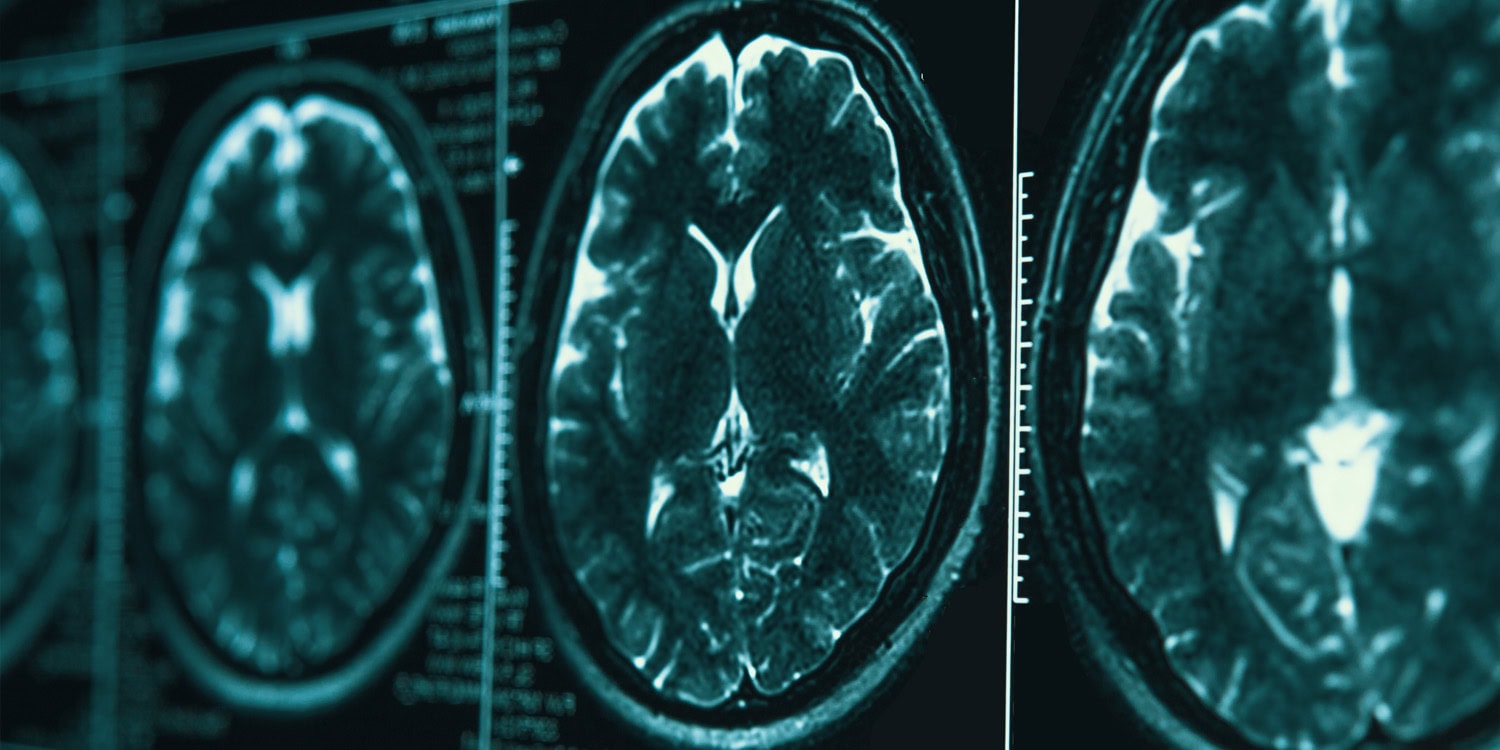
An exploratory study of people with major depressive disorder found that the shape and structure of several regions of the brain’s outer layer, measured before treatment began, were associated with how much patients’ symptoms improved over about eight weeks of taking the antidepressant vortioxetine. The regions included areas of the frontal lobes involved in emotion regulation and planning, visual areas at the back of the brain, and the insula, a region involved in emotional awareness and in sensing the body’s internal state. The paper was published in BMC Psychiatry.
Major depressive disorder (MDD) is a common and often recurring psychiatric condition that can substantially impair everyday functioning and quality of life. Although many antidepressant medications are available, only about one-third of patients achieve remission, meaning their symptoms largely resolve, after their first treatment. People differ considerably in how well they respond to antidepressants, making it difficult to know in advance which treatment will work best for a particular patient.
Researchers are therefore trying to identify characteristics that might help predict treatment response and support more personalized approaches to care. Clinical factors such as the initial severity of depression, duration of illness, anxiety symptoms, and early improvement during treatment have all been linked to later outcomes.
Yingna Li and colleagues at Beijing HuiLongGuan Hospital in China used high-resolution structural magnetic resonance imaging, which captures the brain’s anatomy rather than its activity, along with a technique called surface-based morphometry, to explore whether features of the cortex, the brain’s wrinkled outer layer, could help predict improvement in depressive symptoms after antidepressant treatment.
They also wanted to know whether cognitive improvement is associated with these brain characteristics. The researchers were particularly interested in memory performance. They noted that cognitive problems are common in depression and sometimes persist even after mood symptoms improve. Because cognitive impairments can negatively affect the lives of depressed individuals, finding ways to predict their recovery is also an important goal.
The researchers initially recruited 34 adults between the ages of 18 and 50 who met diagnostic criteria for major depressive disorder, along with 26 healthy individuals for comparison who were matched with the depressed participants on age and sex. All of the patients had experienced at least one previous depressive episode and had not taken antidepressants in the eight weeks before enrolling. Four participants with depression and three healthy participants were excluded because of poor-quality brain scans, mostly due to excessive head movement, and 10 more participants with depression did not return for the follow-up visit. As a result, all analyses were performed on 22 participants with depression and 21 healthy participants.
All participants with depression took a daily 10-milligram dose of vortioxetine. Before and after treatment, they were assessed with the 17-item Hamilton Rating Scale for Depression and the Hamilton Anxiety Rating Scale, two clinician-administered measures of symptom severity, and completed the Repeatable Battery for the Assessment of Neuropsychological Status, a set of brief tests of memory, attention, language, and visual-spatial skills. On average, participants’ depression scores fell from about 27 to about 8 over the course of treatment.
Participants with depression had brain scans at the start of the study and again about eight weeks later, after treatment, while healthy participants were scanned once. However, this study analyzed only the scans taken at the start, because the goal was to see whether brain features measured before treatment were linked to later improvement.
The researchers used these scans to measure four features of the cortical surface: cortical thickness; the gyrification index, which reflects how much of the cortex is tucked into folds rather than exposed at the surface; sulcus depth, the depth of the grooves between folds; and fractal dimension, a measure of how complex and irregular the folding pattern is.
Compared with healthy participants, those with depression showed less folding in the right insula.
Among participants with depression, higher fractal dimension, indicating more complex folding, in the left superior frontal cortex, the right rostral middle frontal cortex, and the right lateral occipital cortex was associated with greater symptom improvement. The two frontal regions are part of the prefrontal cortex, which is involved in emotion regulation and executive control, while the lateral occipital cortex is a visual area. Greater cortical thickness in the insula on both sides of the brain and in the left pericalcarine cortex, part of the primary visual cortex, was also linked to greater improvement.
In contrast, lower fractal dimension in the left cuneus, another visual region, and less folding (a lower gyrification index) in the left lateral occipital cortex were associated with greater improvement. Several of these associations, including those involving the insula and the right lateral occipital cortex, concerned improvement in anxiety scores rather than depression scores.
On the cognitive side, shallower grooves (lower sulcus depth) in the left lateral occipital cortex were associated with greater improvement in delayed memory, the ability to recall information after some time has passed since it was first learned.
To check whether these associations might hold up beyond the specific people studied, the researchers used a technique called leave-one-out cross-validation, in which a statistical model is built using all but one participant and then used to predict the remaining participant’s outcome, with the process repeated for everyone.
In these tests, which also accounted for participants’ age and sex, fractal dimension of the left cuneus was associated with changes in depression scores, though this result was only borderline statistically significant, and fractal dimension of the left superior frontal cortex was associated with changes in anxiety scores. The link between sulcus depth in the left lateral occipital cortex and delayed memory did not reach statistical significance in this test.
“Baseline cortical morphometric features were associated with clinical and cognitive changes following antidepressant treatment in MDD [major depressive disorder],” the study authors concluded. They also added that “these findings are preliminary and should be interpreted with caution, pending replication in independent cohorts.”
The study contributes to the scientific understanding of the links between the brain’s structure and the course of depression during treatment. However, it should be noted that the study was conducted on a very small sample of participants, and the authors acknowledge that because many separate analyses were run, some associations may be false positives and their strength is likely overestimated. Results of larger studies may differ.
All patients received the same drug, so the findings may not apply to other antidepressants, and participants were relatively young. There was also no placebo group, so the study cannot separate improvement caused by the medication from improvement that might have happened for other reasons. Additionally, the design of the study does not allow any causal inferences to be derived from the results.
The paper “Baseline cortical structural features are associated with depressive and cognitive improvement in major depressive disorder” was authored by Yingna Li, Shuping Tian, Siyan Zan, Ruonan Du, Wenzhou Liang, Liying Zhao, and Zhiren Wang.